A tissue-sparing way to lock a painful level in the low back from behind. A small implant with its own locking screw is placed across each facet joint through an opening about 16 mm wide, then paired with an interbody cage at the same level. Dr. Hobbs uses the Providence Medical Technology CORUS-LX™ system, the lumbar counterpart of the DTRAX® cervical technique, for one- and two-level fusions from L4 to S1.
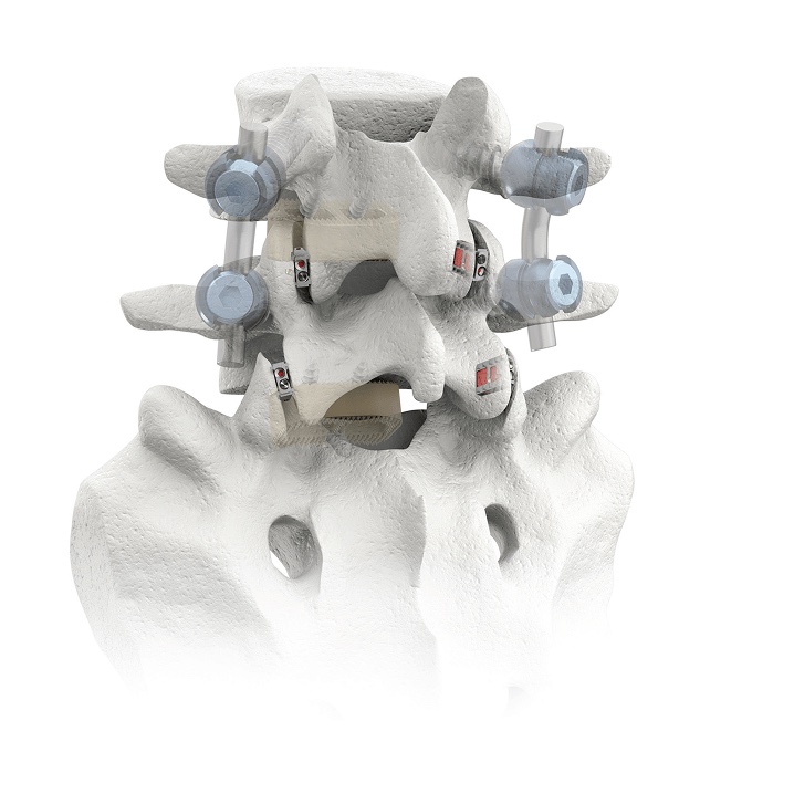
Every level of the low back is joined by three joints: the disc in front and a pair of facet joints at the back. A lumbar fusion stops the painful motion at a level by replacing the disc with a cage and then holding everything still until bone grows across. For decades, “holding it still” has meant pedicle screws and rods placed through a sizable exposure of the back muscles.
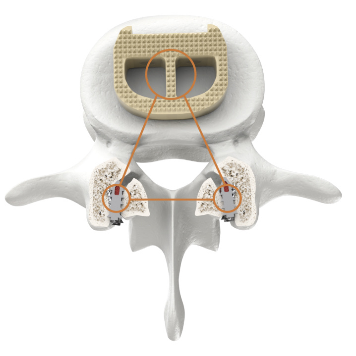
Posterior lumbar facet fusion moves that fixation into the facet joints themselves. Through a mini-open portal no larger than 16 mm on each side, the joint is prepared and a CORUS-LX implant is seated across it. The implant has an integrated self-locking screw and doubles as a reservoir for bone graft, so each facet is fixed and grafted in one step. With a cage in front and an implant in each facet behind, the level is held at three points around the disc.
CORUS-LX is indicated for posterior lumbar fusion in lumbar degenerative disc disease from L4 to S1, as an adjunct to a one- or two-level interbody fusion. It is always paired with an FDA-cleared interbody device at the same level, and it can be used with or without pedicle screws and rods. It is the lumbar relative of the DTRAX cervical system Dr. Hobbs uses in the neck.
Hardware holds only as well as the bone it sits in. A 2021 CT study measured bone density across the lumbar segment and found the facet’s articular processes far denser, and far more cortical, than the vertebral body where cages sit, and denser than the pedicles where screws go. That is the case for anchoring a fusion in the facets.
Mean bone mineral density (mg Ca-HA/mL) and cortical bone fraction by region. Odeh et al., Global Spine Journal, 2021, as reported by the manufacturer.
Tap each step. The same sequence is repeated on the other side of the spine, and at the second level when two are fused.
Simplified illustration for education, courtesy of the manufacturer; not an exact anatomical depiction. Pedicle screws and rods are added when Dr. Hobbs wants additional stability.
A fusion heals when the level stops moving. In a 2025 biomechanical study, cadaver spines were fused at L2–L3 with a lateral interbody cage (LLIF) and then tested three ways: the cage alone, the cage with pedicle screws and rods, and the cage with CORUS-LX facet fixation.
Either form of posterior fixation cut motion sharply compared with the cage alone. Facet fixation matched or bettered the screw-and-rod construct in flexion, extension, and axial rotation, and allowed a little more motion in lateral bending. The authors called it a viable alternative for adding stability to lateral interbody fusion.
This is a cadaver study at one level above the range CORUS-LX is cleared for, and it was presented as a meeting ePoster rather than a peer-reviewed paper. It shows how the construct behaves mechanically, not how patients do.
Average motion at L2–L3, degrees
Lower is stiffer. Values for the CORUS-LX construct are labeled; hover any bar for its value.
| Motion tested | LLIF alone | LLIF + pedicle screws and rods | LLIF + CORUS-LX facet fixation |
|---|---|---|---|
| Flexion | 0.84° | 0.10° | 0.06° |
| Extension | 0.81° | 0.32° | 0.28° |
| Lateral bending | 1.22° | 0.43° | 0.56° |
| Axial rotation | 0.53° | 0.43° | 0.28° |
Values read from the chart published by the manufacturer; approximate to about 0.02°. Zhou et al., “Biomechanical stability of LLIF with posterior intrafacet fixation,” ePoster, Spine Summit 2025. Source.
Facet fixation is a way to hold a fusion, not a reason to have one. Dr. Hobbs considers it for skeletally mature adults who already need a one- or two-level lumbar fusion at L4–S1 and who:
Physical therapy, medication, and usually injections have been tried without lasting relief from degenerative disc disease at the lowest lumbar levels.
Older patients, softer bone, or prior surgery, where anchoring in the densest bone of the segment is an advantage. The clinical data for the system come from high-risk and revision patients.
LLIF and ALIF place the cage from the side or front; facet fixation adds posterior stability without a second large incision in the back. See spinal fusion approaches.
A level that did not heal, or a level next to an old fusion, where Dr. Hobbs wants solid fixation with less muscle disruption. See revision spine surgery.
Each has a job it does best. The right one depends on your bone, your alignment, and how many levels are involved.
| Facet fixation (CORUS-LX) + cage | Pedicle screws and rods + cage | Cage alone | |
|---|---|---|---|
| Where it anchors | The facet joints, the densest bone in the segment | The pedicles, through to the vertebral body | The disc space only |
| Approach | Mini-open portal about 16 mm on each side; muscle spread | Open or percutaneous posterior exposure | No posterior incision |
| Stiffness on the bench | Comparable to screws and rods in flexion, extension, and rotation; a little more motion in lateral bending | The traditional standard | Least; relies on the cage and the patient’s own ligaments |
| Hardware profile | Low; sits inside the joint | Screw heads and rods above the bone | None posteriorly |
| Best for | One- or two-level fusions at L4–S1, including high-risk and revision patients | Deformity, instability, and multilevel constructs | Selected single levels with good bone and alignment |
The approaches are often combined: facet fixation can be added to a screw-and-rod construct, and a cage is always part of the plan. Dr. Hobbs matches the construct to your imaging and bone quality.
Lumbar facet fixation is newer than its cervical cousin, and most of the data so far come from the manufacturer. Here is what has been reported.
Defined as less than 5 degrees of motion on X-ray, in a study of 57 high-risk and revision patients treated with the lumbar facet fixation system as an adjunct to interbody fusion.
Manufacturer study CLN-PMT-145, Providence Medical TechnologyIn the same 57-patient series; 79 percent of patients reported improved pain.
Manufacturer study CLN-PMT-145About 392 versus 147 mg/mL, with roughly five times the cortical fraction, in a CT study of the lumbar segment.
Odeh et al., Global Spine J, 2021In cadaver testing of lateral interbody fusion at L2–L3, across flexion, extension, lateral bending, and axial rotation.
Zhou et al., ePoster, Spine Summit 2025The implants do their job immediately. The bone takes longer. Recovery follows the interbody fusion it supports; the smaller posterior footprint mainly changes the first weeks.
General anesthesia. The facet implants are placed through two small portals, usually in the same operation as the cage. Most patients are walking the same day; the hospital stay depends on the interbody approach and the number of levels.
Walking and light daily activity resume quickly. Bending, lifting, and twisting are limited while the construct protects the level. Soreness at the portals fades over a couple of weeks.
This is when bone bridges the disc space and the facet joints. Activity is increased step by step, therapy rebuilds core strength, and Dr. Hobbs confirms healing on follow-up imaging.
Facet fixation sits alongside the other tools Dr. Hobbs uses to fuse a level with the smallest footprint that solves the problem.

ABNS
Residency
M.D.Facet fixation, pedicle screws, patient-specific cages and rods, and navigation, chosen per patient rather than one construct for everyone.
Specialty-trained in minimally invasive techniques; the same facet-based approach is part of his cervical practice with DTRAX.
Fusion is considered only after nonsurgical care has failed and imaging confirms the level.
Levels that did not heal, or that failed next to an old fusion, are a regular part of his practice.
University of Kentucky medical degree; neurosurgery residency at the University of Chicago, serving as chief resident.
Practicing at Northwest Indiana's most preferred orthopedic and spine practice, with offices in Crown Point & Chesterton.
If a lumbar fusion has been recommended, Dr. Hobbs will review your imaging and explain whether facet fixation, pedicle screws, a combination, or continued conservative care is the right next step for your bone and your alignment.
(219) 250-5010Monday – Friday · 8:00 AM – 4:30 PM
500 E. 109th Avenue
Crown Point, IN 46307
601 Gateway Boulevard
Chesterton, IN 46304
Your next step
Jonathan G. Hobbs, M.D. is a board-certified neurosurgeon at Lakeshore Bone & Joint Institute. For questions about posterior lumbar facet fusion, call the office to arrange an individual evaluation and discuss which options may be appropriate.
Consultation offices: Crown Point, Chesterton. Your symptoms, examination, imaging, and prior treatment help guide the discussion.
Page updated . About content and medical review dates.