Read all five sample findings
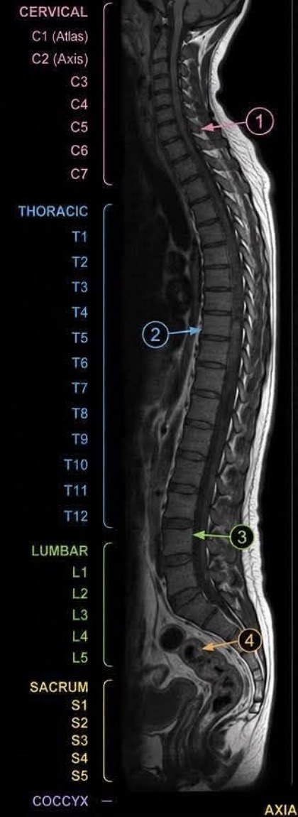
Cervical Spine: C5–C6
Sample report: Posterior disc bulge at C5–C6 indenting the anterior thecal sac. Mild spinal canal stenosis. No significant neural foraminal narrowing. Cervical cord signal is normal.
In plain English: The cushion between the 5th and 6th neck vertebrae is spreading backward and gently pressing on the front of the sac that holds your spinal cord, slightly narrowing the canal. The narrowing is mild, the side doorways where nerves exit are open, and the spinal cord itself looks healthy. This is a very common neck finding.
Herniated disc
Thoracic Spine: T7–T8
Sample report: Small central disc protrusion at T7–T8 mildly indenting the anterior thecal sac. No significant spinal canal stenosis. No neural foraminal narrowing. Thoracic cord signal is normal.
In plain English: In the mid-back, a small focal bit of disc is pushing straight backward and just touching the front of the nerve sac. There is no meaningful narrowing of the canal or the nerve exits, and the spinal cord looks normal. Mid-back disc findings like this are usually quiet and rarely cause symptoms.
Disc protrusion
Lumbar Spine: L4–L5
Sample report: Broad-based posterior disc bulge at L4–L5 with mild ligamentum flavum hypertrophy. Mild to moderate central canal stenosis. Mild bilateral neural foraminal narrowing. No significant spondylolisthesis.
In plain English: In the lower back, the disc has spread evenly backward and the ligament lining the back of the canal has thickened a little. Together they narrow the main canal to a mild-to-moderate degree and slightly tighten the nerve exits on both sides. The vertebrae are still lined up (no slippage). This combination is the classic picture of spinal stenosis.
Spinal stenosis
Lumbosacral Junction: L5–S1
Sample report: Posterior disc bulge at L5–S1 abutting the bilateral traversing S1 nerve roots. Mild bilateral neural foraminal narrowing. No spondylolisthesis.
In plain English: At the very bottom of the spine, the disc is bulging back far enough to touch the S1 nerve roots on both sides as they pass by — without clearly compressing them. The nerve exits are mildly tight. The S1 nerve is the one that runs down the back of the leg, so this is a level that can produce sciatica when it is irritated.
Sciatica
Sacrum & Coccyx: Pelvis
Sample report: Sacroiliac joints appear normal. No acute fracture or marrow edema. Coccyx is normal.
In plain English: The joints where the spine meets the pelvis (the SI joints), the bone itself, and the tailbone all look normal — no fracture, no sign of recent injury or inflammation. A clean bill of health for the bottom of the spine.