A tissue-sparing way to fuse a painful or pinched level in the neck from the back — through two openings about a centimeter wide, without cutting muscle or working past the throat. Dr. Hobbs uses the Providence Medical DTRAX® system for cervical foraminal stenosis, facet arthritis, and anterior fusions that never fully healed.
Each level of the neck is joined by three joints: the disc in front and a pair of small facet joints at the back. When a facet joint wears out, or when the opening beside it narrows and pinches the nerve root to the arm (cervical foraminal stenosis), one option is to stop that joint from moving and let it heal into solid bone — a fusion.
Traditionally that meant either a fusion from the front of the neck (ACDF) or a long incision down the back with the neck muscles stripped off the bone. Minimally invasive posterior cervical fusion reaches the same facet joints from the back through two openings about a centimeter wide. Dr. Hobbs uses the Providence Medical Technology DTRAX® Spinal System: a set of narrow instruments that work between muscle fibers to open the joint, prepare its surfaces, and place a small cage packed with bone graft. The cage props the joint open, which also widens the exit tunnel for the nerve, and over the following months the joint fuses.
The approach does not go near the throat, esophagus, or voice box, and it leaves the front of the spine untouched — which is why it is also used to rescue an anterior fusion that never fully healed and to reinforce high-risk multilevel anterior fusions.
This animation walks through the tissue-sparing posterior approach: reaching the facet joints through narrow instruments, preparing the joint, and placing the cage that props it open while it fuses.
Animation: “Posterior Cervical Fusion Technique.” Educational illustration of the technique — not footage of an actual patient.
Tap each step to see what happens at a single facet joint. The same sequence is repeated on the other side.
Simplified illustration for education — not an exact anatomical depiction. Fixation screws may be added when extra stability is needed.
This is a targeted tool, not an all-purpose neck operation. Dr. Hobbs considers it for skeletally mature adults with degenerative disease between C3 and C7 who have:
Physical therapy, medication, and often an injection have been tried without lasting relief. Surgery is not the first step.
A nerve root pinched in its exit tunnel at the back of the neck. Propping the facet joint open widens that tunnel. See radiculopathy.
Facet arthritis that has been confirmed as the pain source, where fusing the joint stops the painful motion.
A pseudarthrosis after ACDF can be stabilized from behind without reopening the front of the neck. Also used to reinforce high-risk multilevel anterior fusions.
Each approach has a job it does best. The right one depends on what is being compressed and from where.
| Tissue-sparing posterior fusion (DTRAX) | ACDF (from the front) | Traditional open posterior fusion | |
|---|---|---|---|
| Where the incision is | Back of the neck: two openings about 1 cm wide | Front of the neck, in a skin crease | Midline of the back of the neck, several inches |
| Muscle | Spread apart between fibers, not cut | Little muscle, but works past the esophagus and voice-box nerves | Neck muscles stripped from the bone and retracted |
| What it is best at | Foraminal stenosis and facet pain; rescuing a non-healing ACDF | Removing a disc or bone spur pressing the cord or nerve from the front | Instability, multilevel cord compression, deformity |
| Typical stay | Same day or one night in published series | Usually one night | Usually several nights |
| Trade-offs | Cannot remove a disc from the front; not for cord compression | Temporary swallowing trouble or hoarseness is common early on | More postoperative pain and a longer recovery |
Sometimes approaches are combined — for example, a posterior tissue-sparing fusion added behind a multilevel ACDF. Dr. Hobbs matches the approach to your imaging and symptoms.
The DTRAX system received FDA 510(k) clearance in 2018 for posterior cervical fusion in patients with cervical degenerative disc disease, and the tissue-sparing technique has been studied in peer-reviewed series for more than a decade.
In 60 patients treated for single-level cervical radiculopathy, with no reoperations and no device-related complications reported.
McCormack et al., J Neurosurg Spine, 2013Across 150 cases in which the technique was used to revise an anterior fusion that had not healed; no wound infections were reported.
Lemons et al., J Craniovertebr Junction Spine, 2024In a 271-patient device registry, with procedure times of roughly 51 to 88 minutes and blood loss of 32 to 75 mL depending on the number of levels.
Siemionow et al., J Spine Surg, 2018Cages placed in both facet joints from behind increased foraminal area and height in 43 patients, the mechanism behind arm-pain relief.
Siemionow et al., Eur Spine J, 2016The cage does its job immediately. The bone takes longer. Here is how the two timelines fit together.
General anesthesia; the procedure itself typically takes about an hour for one level. Most patients are walking within hours and go home the same day or after one night, with two small dressings at the back of the neck.
Walking and light daily activity resume quickly. Heavy lifting and forceful neck motion are limited while the joint begins to heal. Arm pain from a pinched root often eases early; soreness at the openings fades over a couple of weeks.
This is when bone actually bridges the facet joint. Activity is increased step by step, therapy rebuilds neck strength, and Dr. Hobbs confirms healing on follow-up imaging. Your plan is tailored to the number of levels treated and your health.
Posterior cervical fusion with DTRAX sits alongside the other tissue-sparing tools Dr. Hobbs uses to treat the spine with the smallest footprint that solves the problem.

ABNS
Residency
M.D.Trained specifically in surgery of the spinal cord and nerves, so the choice between decompression and fusion is made by a specialist in both.
Specialty-trained in tissue-sparing techniques; most of his procedures are outpatient.
At least six weeks of nonsurgical care comes before this operation is considered.
Rescuing anterior fusions that did not heal is part of his revision spine practice.
University of Kentucky medical degree; neurosurgery residency at the University of Chicago, serving as chief resident.
Practicing at Northwest Indiana's most preferred orthopedic and spine practice, with offices in Crown Point & Chesterton.
If arm pain, a worn facet joint, or a fusion that never healed has you weighing surgery, Dr. Hobbs will review your imaging and explain whether tissue-sparing posterior fusion, an anterior approach, or continued conservative care is the right next step.
(219) 250-5010Monday – Friday · 8:00 AM – 5:00 PM
500 E. 109th Avenue
Crown Point, IN 46307
601 Gateway Boulevard
Chesterton, IN 46304
Your next step
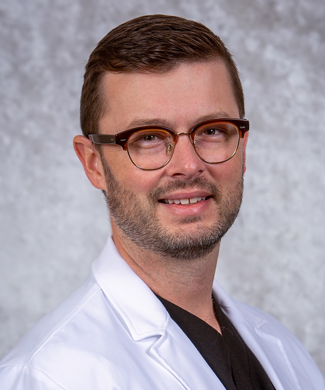
Jonathan G. Hobbs, M.D. is a board-certified neurosurgeon at Lakeshore Bone & Joint Institute. For questions about minimally invasive posterior cervical fusion, call the office to arrange an individual evaluation and discuss which options may be appropriate.
Consultation offices: Crown Point, Chesterton. Your symptoms, examination, imaging, and prior treatment help guide the discussion.
Page updated . About content and medical review dates.