Now accepting new patients Crown Point & Chesterton, Indiana
Home / Procedures / Microdiscectomy & Laminectomy
Decompression Surgery

Microdiscectomy & Laminectomy

Two of the most common and effective procedures for relieving pinched nerves in the spine. Use the interactive guide below to explore how each one works, what it treats, and what recovery looks like — then call Dr. Hobbs with your questions.
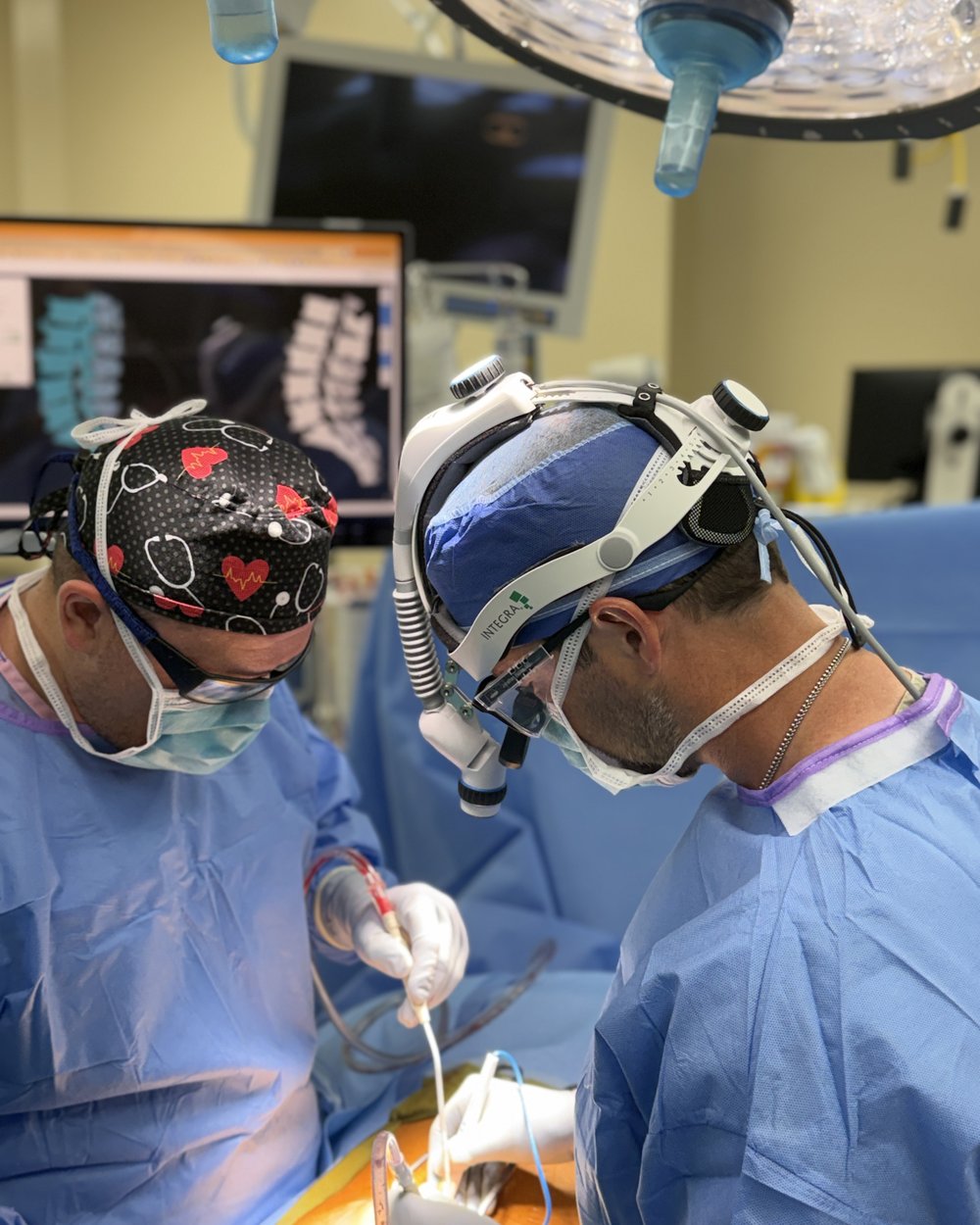
Dr. Hobbs operating with surgical loupes and a headlight while spine imaging is displayed on the monitor behind him
Microscope-assistedDr. Hobbs performing a decompression with magnification and live imaging
Incisions often under one inch. A microscope and tubular retractors spare the surrounding muscle.
Many patients home the same day. Microdiscectomy is usually outpatient; laminectomy often is too.
Surgery is not the first step. Therapy, medication, and injections are tried first whenever it is safe to wait.
Medically reviewed by Jonathan G. Hobbs, M.D. · Updated June 2026
The idea

Relieving Pressure on a Pinched Nerve

Leg pain, sciatica, numbness, and difficulty walking are often caused by something pressing on the nerves in your spine — either a herniated disc or a narrowed spinal canal (stenosis). Microdiscectomy and laminectomy are decompression procedures that remove what's causing the pressure. Both can be performed with minimally invasive, tissue-sparing techniques.

Interactive — choose a procedure

Explore Each Procedure

Each one relieves a different kind of pressure. Tap a procedure to see what it removes and why.

Microdiscectomy

When a disc herniates, its soft inner material pushes out and presses on a nearby spinal nerve — the usual cause of sciatica. In a microdiscectomy, Dr. Hobbs removes just the herniated fragment through a small incision using a microscope, taking the pressure off the nerve while leaving the rest of the disc intact.

Best for
Herniated disc, sciatica, leg pain & radiculopathy
Approach
Small incision (often < 1 inch), microscope-assisted
Anesthesia
General; typically about one hour
Hospital stay
Often same-day / outpatient
Recovery
Light activity in 1–2 weeks; fuller recovery ~6 weeks
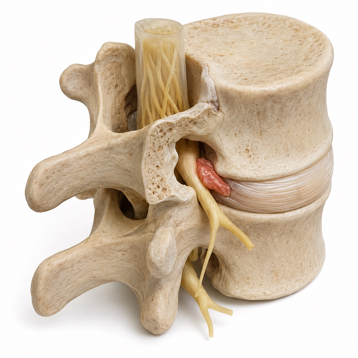
Three-dimensional lumbar spine illustration showing a coral-colored herniated disc fragment beside a yellow nerve root
Herniated disc & nerve rootThe highlighted disc fragment presses against a nearby nerve.

Laminectomy

In spinal stenosis, the spinal canal narrows and squeezes the nerves — often causing leg pain, heaviness, or cramping that worsens with walking. In a laminectomy, Dr. Hobbs removes part or all of the lamina (the bony roof of the canal) to reopen the space and free the nerves, preserving as much normal structure as possible.

Best for
Spinal stenosis, neurogenic claudication, leg pain with walking
Approach
Minimally invasive when possible; removes the lamina
Anesthesia
General; length depends on levels treated
Hospital stay
Outpatient or a short stay, depending on the case
Recovery
Progresses over several weeks; most activity by 6–12 weeks
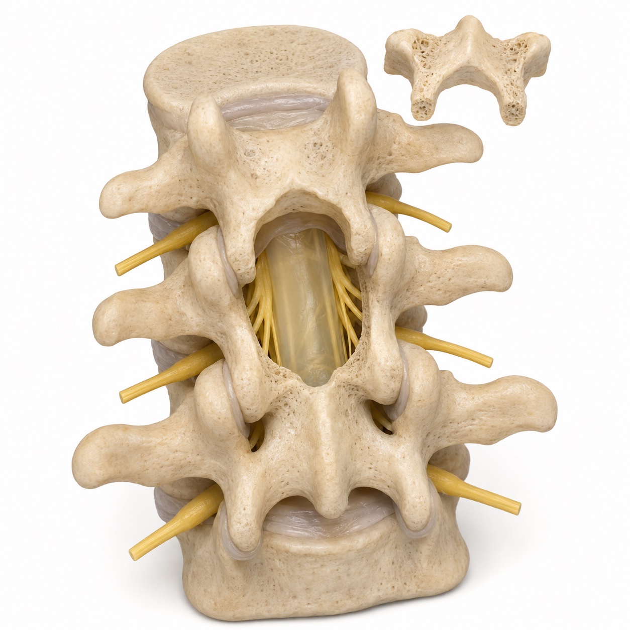
Three-dimensional posterior lumbar spine illustration showing a central opening after laminectomy, with the removed bone displayed above it
Space around the nervesThe removed section of lamina is shown above the opening.

Educational illustrations. Selected structures are cut away or separated to show the anatomy.

Interactive — click each step

Your Procedure, Step by Step

From your first visit to walking out the door, here is how a decompression unfolds.

Compare

Side-by-Side Comparison

Microdiscectomy Laminectomy
Main problemHerniated discSpinal stenosis
What's removedHerniated disc fragmentPart of the lamina (bony roof)
Typical symptom relievedSciatica / leg painLeg pain with walking
IncisionOften < 1 inchSmall; varies by levels
Hospital stayOften same-dayOutpatient or short stay
Fuller recovery~6 weeks~6–12 weeks

Sometimes both are combined — for example, removing a disc fragment and widening the canal in the same operation. Dr. Hobbs tailors the plan to what your imaging and symptoms show.

Interactive — tap a milestone

What Recovery Looks Like

Decompression recovers faster than fusion because nothing has to heal into bone — the nerve simply needs room and time.

Timelines are typical ranges and vary with the procedure, number of levels treated, and individual healing. Dr. Hobbs gives every patient a personalized recovery plan.

Technology

Precision Built In

A good decompression frees the nerve completely while disturbing as little else as possible. Dr. Hobbs pairs magnification and image guidance with muscle-sparing technique to do exactly that.

Candidacy

Are You a Candidate?

Surgery is usually considered after nonsurgical care — rest, medication, physical therapy, and injections — hasn't given enough relief, or when symptoms are progressing. You may be a candidate if you have:

Sudden severe weakness, or loss of bladder or bowel control, can be a surgical emergency — seek care right away or call 911.

When is decompression alone not enough?

If the narrowed level is also unstable or a vertebra has slipped forward, removing bone alone can leave the segment loose. In those cases Dr. Hobbs may pair the decompression with stabilization — motion-preserving when possible, fusion when needed.

Fusion & stabilization
Jonathan G. Hobbs, M.D.
Board-Certified Neurosurgeon · Spine Surgery
American Board of Neurological SurgeryABNS
Certified
The University of ChicagoResidency
Chicago
University of KentuckyM.D.
Kentucky
Why Dr. Hobbs

Why Choose Dr. Hobbs for Decompression Surgery

  • Minimally Invasive Focus

    Tissue-sparing, microscope- and navigation-guided techniques for smaller incisions and faster recovery.

  • Image-Guided Precision

    StealthStation navigation confirms the level and guides an accurate, targeted decompression.

  • Board-Certified Neurosurgeon

    Certified by the American Board of Neurological Surgery.

  • Elite Training

    University of Kentucky medical degree; neurosurgery residency at the University of Chicago, serving as chief resident.

  • Conservative-First Philosophy

    Nonsurgical options are explored first; surgery is recommended only when it is the right answer.

  • Lakeshore Bone & Joint Institute

    Practicing at Northwest Indiana's most preferred orthopedic and spine practice, with offices in Crown Point & Chesterton.

Frequently Asked Questions About Microdiscectomy & Laminectomy

Both relieve pressure on the spinal nerves, but they target different problems. A microdiscectomy removes the herniated portion of a disc that is pinching a nerve — most often the cause of sciatica. A laminectomy removes part of the lamina, the bony roof of the spinal canal, to create more room for nerves squeezed by spinal stenosis. Dr. Hobbs chooses the procedure based on what is compressing your nerves.
A microdiscectomy is often performed on an outpatient basis, meaning most patients go home the same day. It is one of the most common and successful minimally invasive spine procedures, and many patients experience immediate relief of their leg pain after surgery.
Recovery varies by procedure and individual. After a microdiscectomy, many patients return to light activity within 1-2 weeks and recover more fully over about 6 weeks. After a laminectomy, recovery generally progresses over several weeks, with most normal activities resumed within 6 to 12 weeks. Dr. Hobbs provides an individualized recovery plan for every patient.
Dr. Hobbs uses minimally invasive techniques whenever appropriate. A microdiscectomy is often performed through an incision of about one inch or less. A laminectomy may require a slightly larger incision depending on how many levels are treated, but minimally invasive approaches preserve more muscle and tissue and leave smaller scars than traditional open surgery.
Surgery is usually considered after conservative treatments such as medication, physical therapy, and injections have not provided adequate relief, or when there is progressive weakness or numbness. Good candidates typically have leg pain, sciatica, or difficulty walking caused by a herniated disc or spinal stenosis confirmed on imaging. Dr. Hobbs evaluates each patient individually.
Yes. Jonathan G. Hobbs, M.D. is a board-certified neurosurgeon who performs microdiscectomy and laminectomy using minimally invasive, tissue-sparing techniques with image-guided navigation. He practices at Lakeshore Bone & Joint Institute, with offices in Crown Point & Chesterton, Indiana.

Get Answers About Your Back or Leg Pain

If sciatica or leg pain is holding you back, find out whether a microdiscectomy or laminectomy could help. Dr. Hobbs will review your imaging and explain your options. Call today to schedule a consultation.

(219) 250-5010

Monday – Friday · 8:00 AM – 4:30 PM

Crown Point Office

500 E. 109th Avenue
Crown Point, IN 46307

Chesterton Office

601 Gateway Boulevard
Chesterton, IN 46304

Your next step

Discuss your spine care with Dr. Hobbs

Jonathan G. Hobbs, M.D. is a board-certified neurosurgeon at Lakeshore Bone & Joint Institute. For questions about microdiscectomy & laminectomy, call the office to arrange an individual evaluation and discuss which options may be appropriate.

Consultation offices: Crown Point, Chesterton. Your symptoms, examination, imaging, and prior treatment help guide the discussion.

Request a free second-opinion review

Page updated . About content and medical review dates.